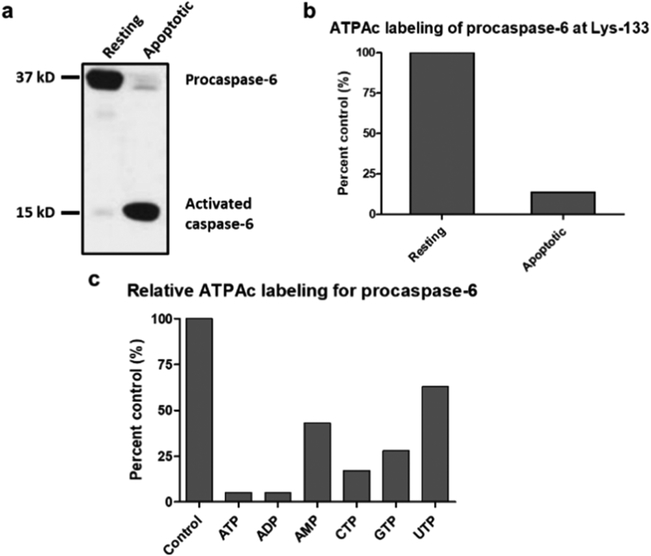
Figure 2.
(a and b) ATPAc labeling of resting and apoptotic Jurkat cell lysates and (c) inhibition of labeling in resting cells by addition of nucleotides. (a) Protein immunoblots of procaspase-6 and caspase-6 in lysates from resting and apoptotic Jurkat cells (induced by anti-FAS antibody 4C3). (b) Both lysates were treated with ATPAc and subjected to our chemoproteomic platform, and the acylated K133 peptides were identified and quantified. The resting lysate containing predominantly procaspase-6 was defined as 100% acylation. (c) Lysates from resting Jurkat cells were individually pretreated (15 min.) with the stated nucleotides (5 mM) and then subjected to the described procedures to quantify the inhibition of formation of the acylated K133 peptide.

