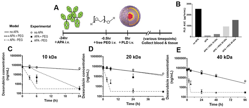
Figure 1: High MW free PEG rescues the pharmacokinetics of PEG-drugs in mice with APA.

(A) We developed a mouse model of APA-mediated ABC by administering defined amounts of APA to naïve mice i.v., followed 24 h later by i.v. injection of PLD ± free PEG. (B) The AUC0-96h of PLD in mice that received the various sizes of free PEG polymer. (C–E) The PK of PLD was measured in naïve mice (open circle) as well as passively immunized mice receiving either i.v. PBS (filled triangle) or various free PEG interventions (filled square), including (C) 10 kDa (2200 mg/kg), (D) 20 kDa (550 mg/kg), and (E) 40 kDa (550 mg/kg) free PEG 30 min prior to PLD administration. Lines depict PK predictions from mPBPK model. For comparison, converting the above mg/kg doses of PEG 10 kDa, 20 kDa, and 40 kDa to molar doses equals 220 μmole/kg, 27.5 μmole/kg, and 13.75 μmole/kg, respectively.
