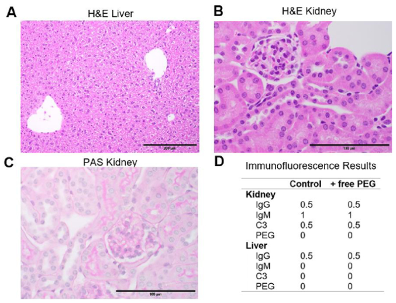
Figure 3: Repeat dosing of High MW free PEG does not cause immune complex deposition or apparent tissue damage.

PEG-sensitized mice were given six weekly injections of 40 kDa free PEG. (A and C) Routine H&E staining of liver (200X magnification) and kidney (600X). Tissue histomorphology is within normal limits. (A) Regionally, hepatocytes in the liver display mild vacuolar change consistent with normal glycogen storage. (B) H&E of the kidney suggests that renal tubular epithelium occasionally display attenuated cells, consistent with post-mortem change, and lack vacuolation. (C) PAS staining of kidney (600X) highlights thin, positive-staining basement membranes of the glomerulus and renal tubules. Note that in the inner lumen of renal tubules there is irregular PAS-positive staining of PAS-positive secretions that are not basement membranes. (D) Summary immunofluorescence (IF) averages for PEG-sensitized mice that received either PBS or free PEG for six weeks. See complete panel of representative IF images in Supplemental 4.
