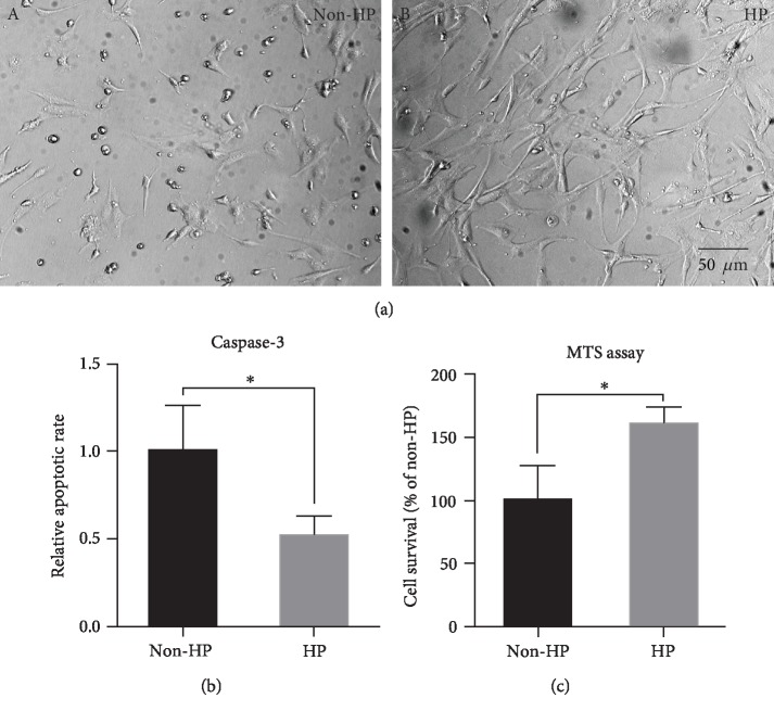
Figure 4.
HP reduced the apoptosis of CV-MSCs after ischemic stimulus. Representative microphotographs taken by light microscopy (a), caspase-3 activity (b), and MTS assay (c) of CV-MSCs with HP or CV-MSCs with non-HP after ischemic stimulus for 24 h. Data are expressed as mean ± standard deviation: ∗p < 0.05 (n = 4).

