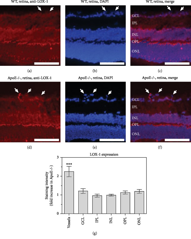
Figure 5.
Immunostainings for the ox-LDL receptor, LOX-1, in retinal cross-sections from wild-type (a–c) and ApoE-/- mice (d–f), respectively. Staining intensity was increased in blood vessels from ApoE-/- mice (g) but did not differ in any of the retinal layers between both genotypes. GCL: ganglion cell layer; IPL: inner plexiform layer; INL: inner nuclear layer; OPL: outer plexiform layer; ONL: outer nuclear layer. Values are presented as mean ± SE (n = 8 per genotype; ∗∗∗P < 0.001). Scale bar = 100 μm.

