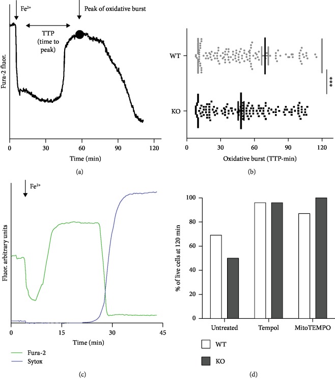
Figure 2.
Single-cell analysis in GSH-depleted MEF cells. (a) The acute Fe2+ entry was followed kinetically at single-cell level in fura-2-loaded MEFs: pyrithione-mediated Fe2+ entry promoted fura-2 fluorescence quenching (excitation at 360 nm, the Ca2+-insensitive wavelength) followed, after a variable period of time, by fluorescence recovery due to oxidation of Fe2+ to Fe3+ likely induced by Fenton reaction. The interval between Fe2+ entry and the peak (indicated by a dot in the representative curve) of oxidative burst (TTP: time to peak) occurred significantly early in KO compared to WT MEFs, as described by the graph in (b); ∗∗∗p < 0.001. (c) The fura-2 dequenching phase can be followed by a loss of fura-2 fluorescence signal (observable also in (a)) as a consequence of an increase in plasmalemma permeability also documented by a concomitant Sytox blue nuclear staining. (d) The percentage of live MEFs (negative for Sytox staining and still loaded with fura-2) at the end of the experiments (120 minutes after acute iron overload) was evaluated, by considering all cells analyzed, under untreated conditions or after pretreatment with 100 μM Tempol and 100 μM MitoTEMPO, cytosolic and mitochondrial ROS scavengers, respectively. Both treatments completely prevented cell death.

