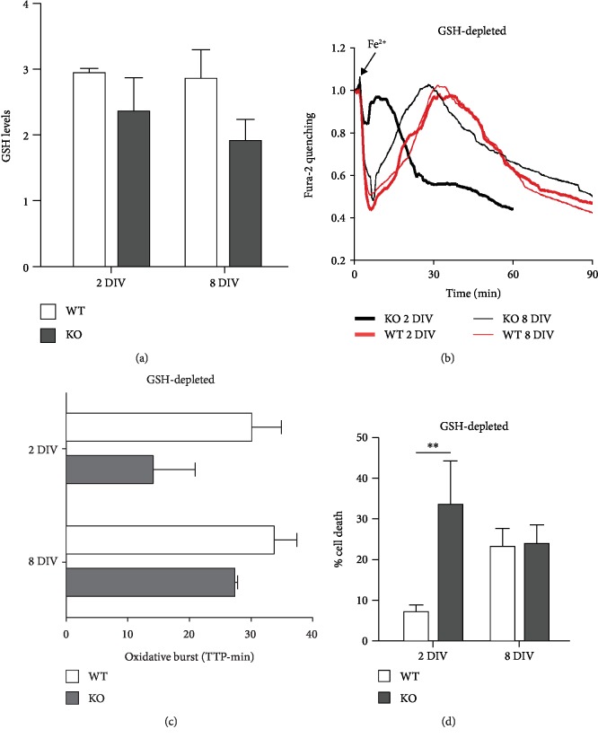
Figure 4.
Role of glutathione in the protection of KO cortical neurons from oxidative stress. (a) During aging in culture, GSH content, analyzed by mBCl as described in Figure 1(c), remained at comparable levels in WT neurons, while it showed a decreasing trend in KO neurons, indicating a minor role for GSH in the establishment of protective mechanisms. (b, c) Glutathione depletion (by O/N treatment with BSO, 300 μM) speeds up all cellular processes following Fe2+ entry (compared with the 2 graphs in Figures 3(a) and 3(b)), but still, 8 DIV KO neurons are more resistant than the 2 DIV KO ones. This was confirmed by the kinetics of oxidative burst (evaluated as in the previous two figures), therefore indicating no major role of GSH in antioxidant protective mechanism. (d) Under conditions of glutathione depletion (as in (b) and (c)), neuronal death was significantly higher in 2 DIV KO compared to WT neurons, while it was similar in 8 DIV neurons; ∗∗p < 0.01.

