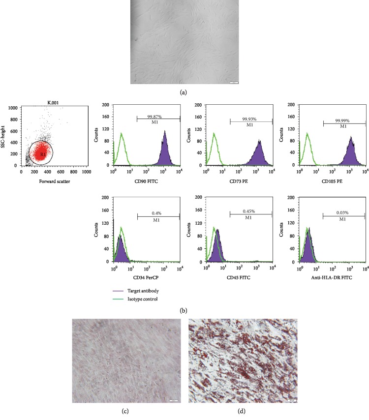
Figure 1.
Identification of human UC-MSCs. (a) Morphological features. The MSCs appeared spindle-like and fibroblastoid-shaped. Scale bar = 100 μm. (b) Flow cytometric analysis of the expression of cell surface markers related to human UC-MSCs. The expression of each antigen was presented with the corresponding isotype control. (c) Alizarin Red S staining of cultured osteogenic human UC-MSCs. Scale bar = 100 μm. (d) Oil Red O staining of cultured adipogenic human UC-MSCs. Scale bar = 50 μm.

