
Figure 2.
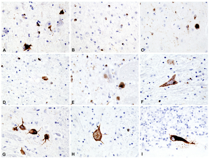
Higher power view of CNS areas stained with pTDP‐43. A. Primary motor cortex. B. White matter adjacent to primary motor cortex. C. Putamen. D. Subthalamic nucleus. E. Substantia nigra. F. Hypoglossal nucleus. G. Inferior olivary nucleus. H. Lumbar spinal cord, anterior horn. I. Cerebellum, vermis (pTDP‐43, 40x original magnification).