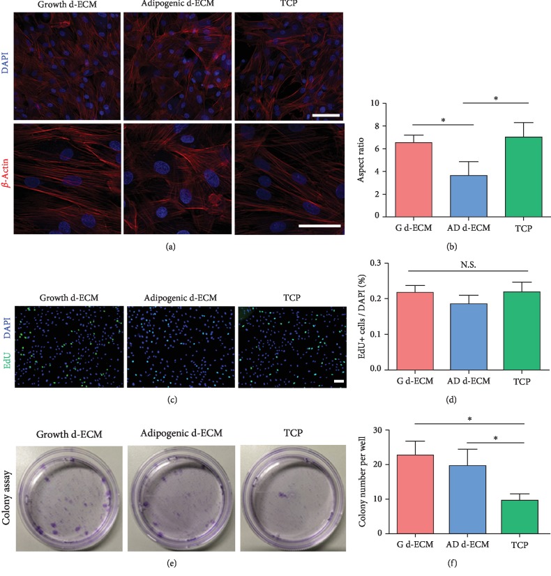
Figure 3.
Cellular morphology and proliferation of ASCs cultured on different substrates. (a) Cellular morphology of ASCs cultured on different substrates. ASCs were reseeded on three different substrates: growth d-ECM, adipogenic d-ECM, and tissue culture polystyrene (TCP). Immunostaining for F-actin. (b) ASC morphology is quantitatively compared by measuring cell aspect ratio using ImageJ software. (c) EdU assay for proliferation of ASCs. (d) Quantitative analysis of EdU+ cell proliferation rate. (e) Colony-forming assay. (f) Quantitative analysis of colony number per well. Results are presented as the mean ± SD; one-way ANOVA followed by Bonferroni's post hoc test analysis for multiple comparison. Scale bar = 100 μm.

