Figure 1.

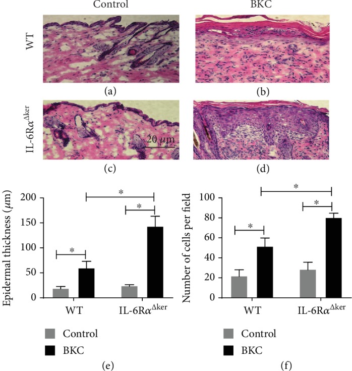
IL-6RαΔker mice present with increased epidermal hyperplasia following irritant exposure. Loss of the IL-6Rα in keratinocytes promotes epidermal hyperplasia during irritant contact dermatitis. WT and IL-6RαΔker mice were exposed to BKC or control for seven (7) consecutive days to induce ICD. 24 hours after irritant exposure, 8 mm biopsies of lesional skin were collected and embedded in O.C.T. compound for histological analysis. Skin samples were cross-sectioned and then hematoxylin and eosin (H&E) stained. Representative H&E stains from WT (a, b) and IL-6RαΔker (c, d) are shown. Quantification of epidermal thickness (e) and cells per field (f) as determined by ImageJ (NIH) is presented. Data are mean ± SD. ∗Significantly different from WT (p ≤ 0.05, n = 15 mice/treatment/genotype).
