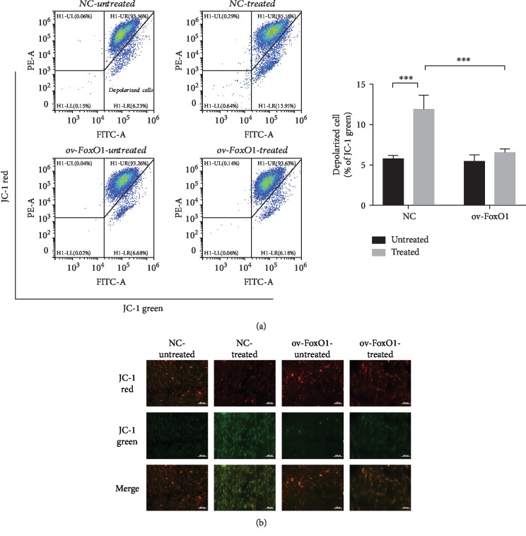
Figure 2.
FoxO1 overexpression improved the mitochondrial membrane potential in hPDLSCs. PDLSC-pMSCV-empty (NC) and PDLSC-pMSCV-FoxO1 (ov-FoxO1) were labeled with JC-1 to determine their mitochondrial membrane potential following treatment with α-MEM or TNF-α (10 ng/mL) for 72 h (a). Quantification of depolarized cells presented as the percent of the total cells. Confocal microscopic images of hPDLSCs were labeled with JC-1 (b). JC-1 aggregates exhibited red fluorescence, and JC-1 monomers exhibited green fluorescence. ∗∗∗p < 0.001.

