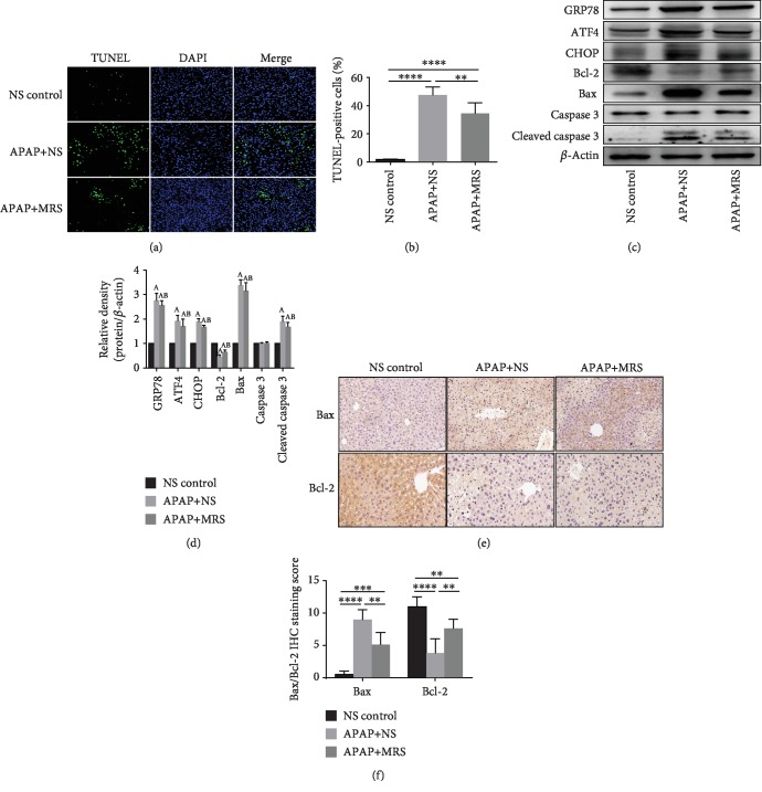
Figure 4.
MRS inhibited ERS-induced apoptosis after APAP challenge. (a) TUNEL fluorescence staining (green), nuclear counterstaining (blue), and merging of both channels in the representative liver sections (200x); (b) percentage of TUNEL-positive cells; (c, d) western blot results of GRP78, ATF4, CHOP, Bcl-2, and Bax expressions in the liver; (e) IHC staining (200x) showing Bax and Bcl-2 expressions in the liver; and (f) relative IHC staining scores (∗p < 0.05, ∗∗p < 0.01, ∗∗∗p < 0.001, and ∗∗∗∗p < 0.0001).

