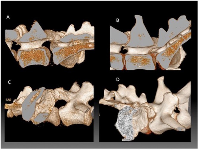
Figure 1.
Three-dimensional reconstructions from CT data showing the lumbosacral junction of a German shepherd dog during flexion compared to extension. The vertebral column has been sectioned in the midline to only include the right half. (A) external detail of the LS junction in flexion, (B) internal detail of the LS junction in flexion, (C) external detail of the LS junction in extension, (D) external detail of the LS junction in extension.

