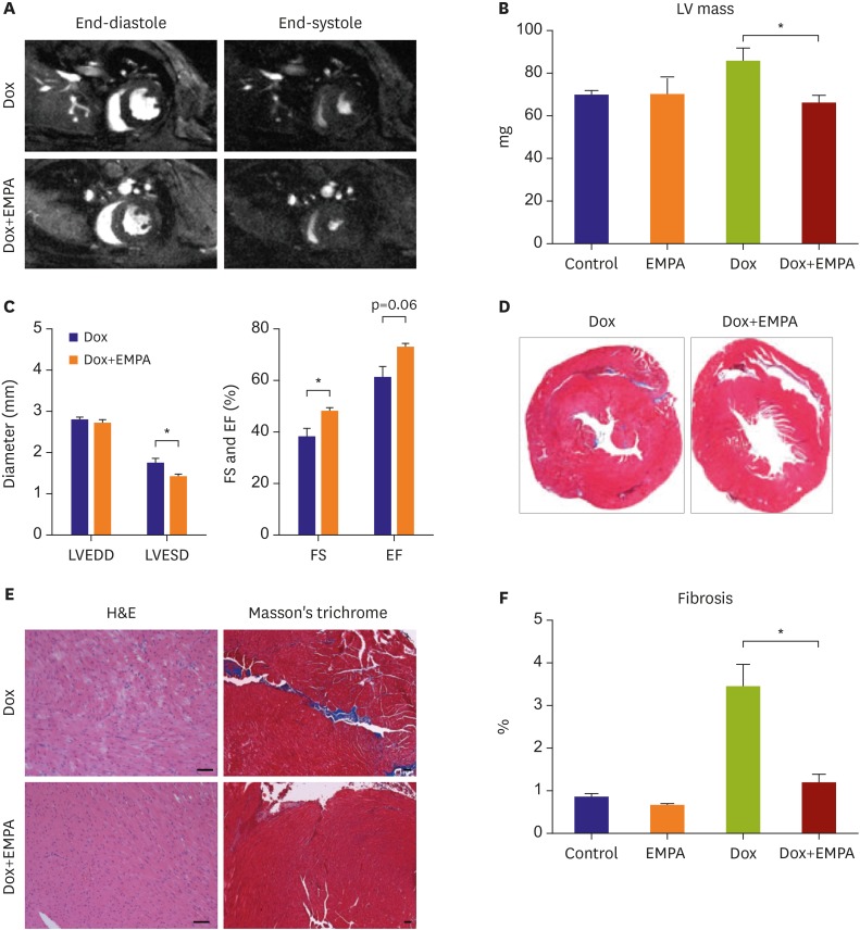
Figure 1. The SGLT2 inhibitor protects against Dox-induced cardiac toxicity. (A) Representative images of Dox-mediated changes in cardiac structure and function from cardiac MRI. (B) and (C) The Dox+EMPA group showed reduced LV mass, decreased LVESD, and improved FS compared with the Dox group. (D) Masson's trichrome stained cross-sections of mice, 2 weeks after single Dox injection. (E) Representative images of LV sections stained with H&E and with Masson's trichrome. The Dox+EMPA group showed less myocardial damage. (F) Quantification of interstitial fibrosis. Scale bar=50 μm. Each bar represents mean±standard error of mean.
Dox = doxorubicin; EF = ejection fraction; EMPA = empagliflozin; FS = fractional shortening; H&E = hematoxylin and eosin; LV = left ventricle; LVEDD = left ventricular end diastolic diameter; LVESD = left ventricular end systolic diameter; MRI = magnetic resonance imaging; SGLT2 = sodium-glucose co-transporter 2.
*p<0.05.

