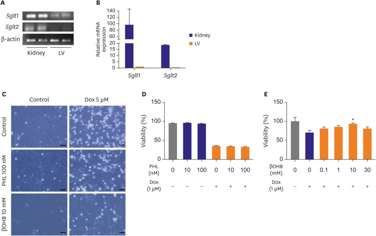
Figure 2. The SGLT inhibitor protects cardiomyocytes by increasing βOHB. (A) and (B) Sglt1 and Sglt2 mRNA expression in mouse LV and kidney by qRT-PCR. (C) Representative light microscopic images of H9C2 cells. H9C2 cells were stimulated with 5 μM Dox or pre-treated with PHL or βOHB for 2 hours and then treated with 5 μM Dox for 24 hours. (D) H9C2 cell viability of the PHL pre-treatment group by an MTT assay. (E) H9C2 cell viability of the βOHB pre-treatment group by an MTT assay. βOHB group showed improved cell viability under 1 μM Dox-induced cardiotoxicity compared to non-treated group. Scale bar=50 μm. Each bar represents mean±standard error of mean.
βOHB = beta hydroxybutyrate; Dox = doxorubicin; LV = left ventricle; mRNA = messenger RNA; MTT = 3-(4,5-dimethylthiazol-2-yl)-2,5-diphenyltetrazolium bromide; PHL = phloridzin; qRT-PCR = quantitative real-time reverse transcription polymerase chain reaction; SGLT = sodium-glucose co-transporter.
*p<0.05 vs. Dox-treated group.

