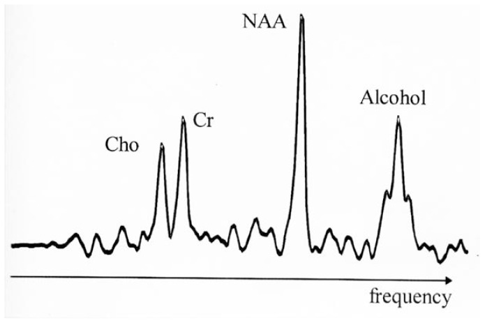
Figure 2.
Magnetic resonance spectrum from hydrogen nuclei in a human brain (the signal from water, which would be much larger than the others, is suppressed). The peaks shown are from the three major signals visible by MRS1 in the normal brain, representing Cho,2 Cr,3 and NAA.4 The magnetic resonance spectrum was obtained after the subject had consumed alcohol; therefore, a fourth peak appears that originates from alcohol in the brain. This peak exhibits the typical triplet structure of alcohol and resonates at alcohol’s characteristic frequency at the right side of the spectrum.
1MRS = magnetic resonance spectroscopy; 2Cho = choline-containing compounds; 3Cr = creatinine-containing compounds; 4NAA = N-acetylaspartate.

