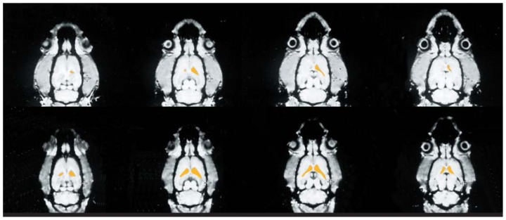
Figure 3.
Small animal magnetic resonance images of a rat brain before and after thiamine deficiency. The images represent horizontal cross-sections progressing from the top of the head (left images) to the base of the skull (right images). The images on the top show the rat’s brain before the experiment; those on the bottom were obtained after the rat received a thiamine-deficient diet for 6 weeks. In each of the cross-sections analyzed, the ventricles (indicated in yellow) were larger after thiamine deficiency than before the experiment.
SOURCE: Adapted from Pentney. R.J.; Alletto, J.J.; Acara, M.A.; Dlugos, C.A.; and Fiel, R.J. Small animal magnetic resonance imaging: A means of studying the development of structural pathologies in the rat brain. Alcoholism: Clinical and Experimental Research 17(6): 1301–1308.1993.

