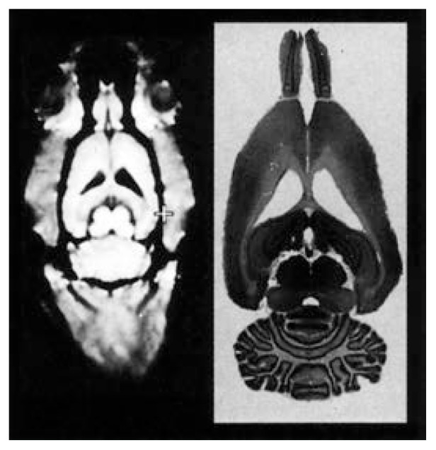
Figure 4.

Comparison of a small animal magnetic resonance (SAMRI) image (left) and the histological examination of the corresponding brain slice (right). The SAMRI image shows the brain as well as the surrounding skull. All the brain structures that can be seen in the histological slice are visible in the corresponding image.
SOURCE: Adapted from Pentney et al. 1993.
