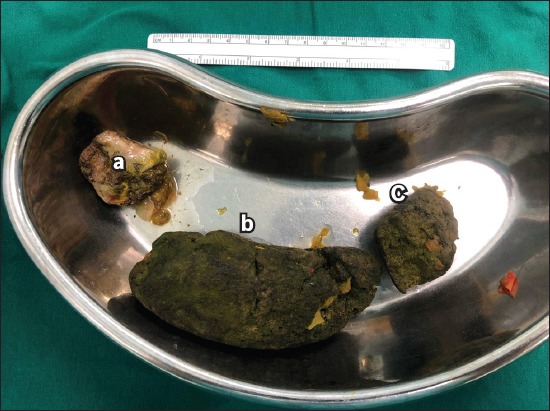
Fig. 1.

Photograph shows gross specimens of a 5-cm phytobezoar obstruction in the distal small bowel retrieved through an enterotomy and primary repair (a & c). Another 15 cm × 6 cm sausage-shaped phytobezoar (b) was also imaged and retrieved from the stomach via gastrotomy and primary repair.
