
Figure 2.
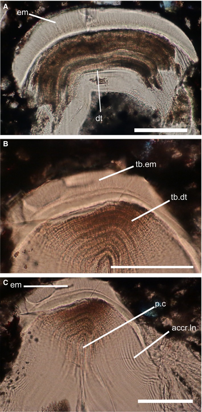
Histological sections through the superficial layer of Astraspis dermoskeleton. (A) Odontode showing the two layers composing the superficial layer. (B) Zoom on the enameloid layer. (C) Zoom on the dentine layer, showing the pulpar cavity. acc.ln, accretion lines; dt, dentine; em, enameloid; p.c, pulpar cavity; tb.dt, dentine tubule; tb.em, enameloid tubule. Scale bars: 100 μm.