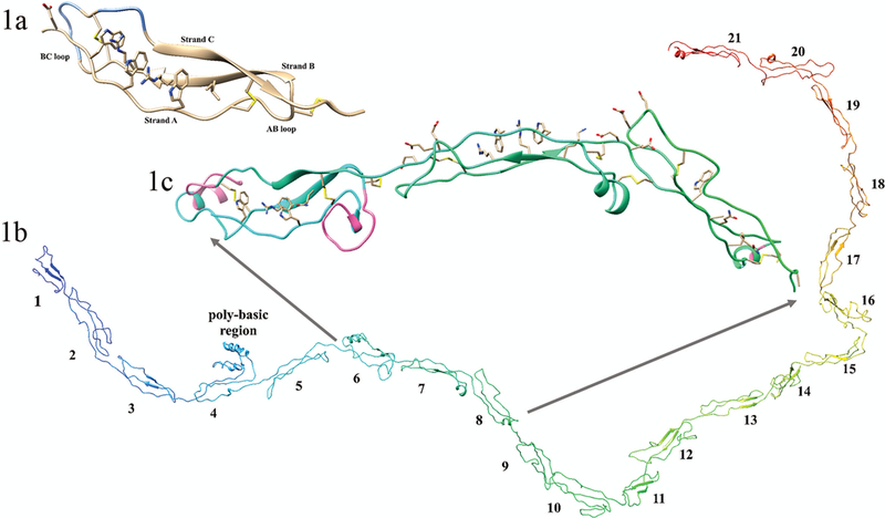
FIGURE 1. Structural features of the second thrombospondin repeat in Thrombospondin-1, and THSD7A.

(1a) Crystal structure image of the second TSR repeat in thrombospondin (created using pdb ID 1LSL). This TSP-1-like domain has 3 strands; strand A, demonstrating the characteristic ripple, and strands B and C showing the characteristic short beta sheets, and the AB and BC loops are indicated. The regions of the BC loop ribbon colored blue are the jar handles. Shown is the Trp-ladder with three Trp residues and two Arg residues and an N-terminal hydrophobic substitution. The disulfides bonds show the C1-C5, C2-C6, and C3-C4 bonding pattern of the TSP-1-like domain. (1b) complete homology of the 21 extracellular TSR domains of THSD7A; N-terminus (blue) and C-terminus (red). Domain numbers are shown for each individual TSR domain. (1c) Expanded image of domains 6 through 8 of THSD7A. Shown are the disulfide bonds of consecutive TSP-1-like (domains 6 and 8) interrupted by a F-spondin-like domain (domain 7) in THSD7A. In domain 6 important structural features of note are the AB loop insertion, the jar handle insertion (insertions shown in pink), and the WxxxxW Trp-ladder. In domain 7 the Trp-ladder comprising three positively charged residues and two Trp residues is demonstrated. Both domains 7 and 8 have negatively charged residues in the BC loop. Domain 8 has a relatively small BC loop insertion (shown in pink) and no Trp-ladder forming cation-pi interactions.
