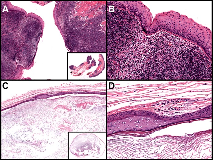
Figure 4.
A and B, Branchial cleft cyst: nonkeratinizing squamous epithelium-lined cysts with abundant lymphocytic aggregates within the cyst wall (A: ×40 [inset: whole slide], B: ×200). C and D, Epidermoid inclusion cyst: thin-walled cysts with a keratinized squamous epithelium lining containing a granular layer; the cyst contains abundant keratin debris or laminated keratin (C: ×40 [inset: whole slide], D: ×200; hematoxylin and eosin staining).

