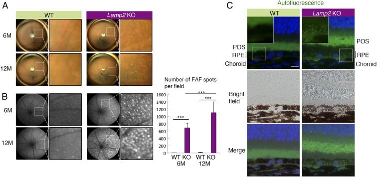
Fig. 2.
Age-dependent fundus pigmentary and autofluorescence abnormality in Lamp2 KO mice. (A and B) Color fundus photography and fundus autofluorescence (FAF) in Lamp2 KO mice and WT mice. WT mice showed normal fundus appearance while Lamp2 KO mice demonstrated atrophic fundus with patchy pigmentary abnormality. Lamp2 KO mice exhibited an age-dependent increase in punctate high-intensity spots in FAF. The numbers of FAF punctate per eye were quantified. n = 6 mice per group. ***P < 0.001. One-way ANOVA with post hoc Tukey HSD test. (C) Representative image of autofluorescence obtained in a setting similar to that for clinical FAF (488-nm excitation) in the paraffin sections of 6-mo-old WT or Lamp2 KO mouse eyes. (Scale bar: 10 μm.) Values are expressed as mean ± SD.

