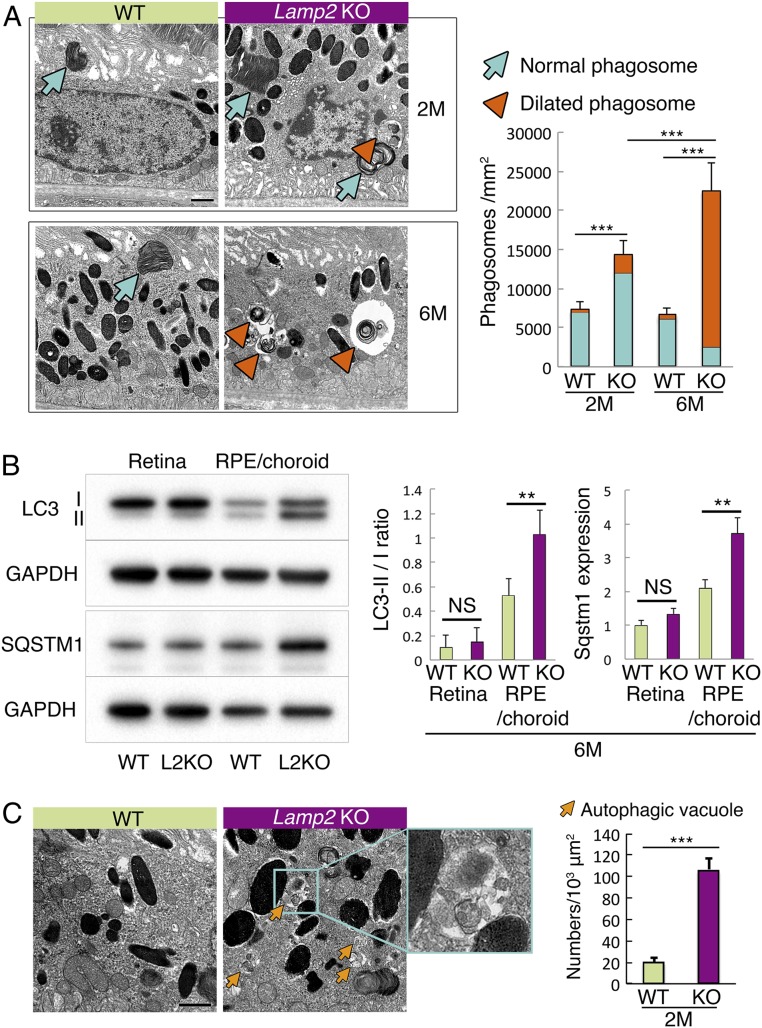
Fig. 6.
Dysregulated autophagic and phagocytic degradation in the LAMP2-deficient RPE. (A) TEM showed increased numbers of phagosomes containing POSs (arrows: normal phagosome; arrowheads: dilated phagosome) in Lamp2 KO mice compared to WT mice. Note the abnormal morphology of dilated phagosomes in 6-mo-old Lamp2 KO mice (arrowheads). The numbers of phagosomes containing identifiable multilayered membranes (disk structure) of POSs were determined at 6 defined regions and mean values were plotted. n = 6 mice per group. ***P < 0.001. One-way ANOVA with post hoc Tukey HSD test. (B) Western blot analyses of LC3 and SQSTM1 in the retina or the RPE/choroid from WT or Lamp2 KO mice. LC3-II/I ratio and SQSTM1 expression of the RPE/choroid was significantly increased in Lamp2 KO mice compared to WT mice. n = 6 eyes from 3 mice per group. **P < 0.01. One-way ANOVA with post hoc Tukey HSD test. (C) Representative TEM images of autophagic vacuoles (arrows) in the RPE of WT and Lamp2 KO mice. Autophagic vacuoles that did not contain identifiable POS disk structures were counted in 6 defined regions, and mean values were plotted. n = 6 mice per group. **P < 0.001, ***P < 0.0001. NS, not significant. Student t test. Values are expressed as mean ± SD. (Scale bars: 1 μm in A and C.)

