Fig. 7.
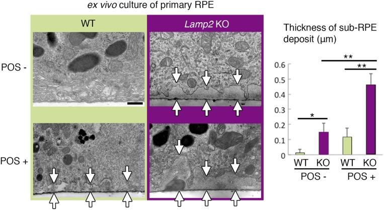
Ex vivo cultures of the LAMP2-deficient RPE recapitulated the sub-RPE deposits. Primary RPE monolayers were isolated as sheets from WT or Lamp2 KO mice and cultured in the presence or absence of POS administration. TEM images of primary RPE cultures on the membrane inserts were obtained. The maximum thickness of basolateral extracellular material was determined at 6 regions per membrane (n = 4 membranes per group). RPE cultures from Lamp2 KO mice showed a significantly increased accumulation of sub-RPE deposits compared to those from WT mice, which became more pronounced with the addition of POSs (arrows indicate the thickness of sub-RPE deposits). *P < 0.05, **P < 0.01. One-way ANOVA with post hoc Tukey HSD test. Values are expressed as mean ± SD. (Scale bar: 0.5 μm.)

