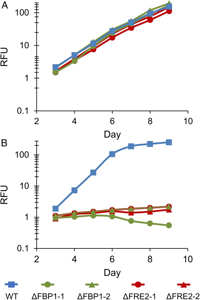
Fig. 1.
Growth of WT, ∆FRE2, and ∆FBP1 P. tricornutum cells in low-iron Aquil media. Relative fluorescence unit (RFU) of diatom cultures grown under 24-h illumination in Aquil media with 10 nM total Fe. Error bars represent ±1 SD of biological triplicate cultures and are obscured by line markers. (A) 100 µM EDTA; (B) 100 µM EDTA and 100 nM DFOB. Two cell lines for each knockout are included, and descriptions of these lines are found in SI Appendix, Table S1.

