Figure 2.
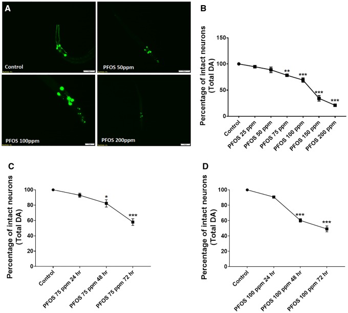
PFOS treatment causes DA cell loss that is concentration and time dependent. Treatment of worms with PFOS (exposure level range: 25–200 ppm for 72 h) resulted in distinct morphological changes such as axon breaks or loss of dendrites, swelling, and loss of soma, representative of neuronal damage in worms (A). The percentage of neuronal loss calculated with respect to total neurons (B). Time course studies at 75 ppm (C) and 100 ppm (D) exhibited a significant impact of exposure time on DA cell loss in worms. Data are presented as mean ± S.E.M. The percentage of intact neurons was calculated by counting the total number of neurons in each worm for 20 worms per experimental group. Data were analyzed using one-way ANOVA followed by Dunnett’s post hoc test. *p < .05, **p < .005, and ***p < .001 (n = 3). Scale bar represents 50 μM (A; Control and 50 ppm PFOS) and 20 µM (PFOS 100 and 200 ppm).

