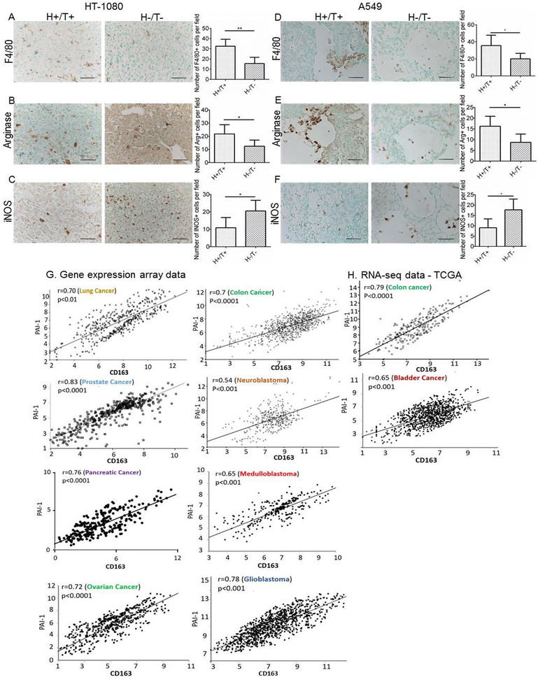
Figure 1. Decrease in macrophage infiltration and arginase expression, and increase in iNOS expression in the absence of host- and tumor-derived PAI-1 in HT-1080 and A549 tumors.
A-F. HT-1080 and A549 cells stably expressing PAI-1 shRNA (T−) or scramble control shRNA (T+) were injected subcutaneously into the right flank of Rag-1−/− PAI-1−/− mice (H-) and Rag-1−/− PAI-1+/+ mice (H+), respectively. Representative immunohistochemical staining of HT-1080 (A-C) and A549 (D-F) tumor samples in the two experimental groups (H+/T+, H−/T−) for F4/80 (A and D), arginase (B and E), and iNOS (C and F); scale bar = 50 μm. The data represent the mean (± SD) of positive cells per section (HT-1080: group H+/T+, n = 5; group H−/T−, n = 5; A549: group H+/T+, n = 5; group H−/T−, n = 4). Two-sided Student t test was applied for comparisons between the two groups. (*p<0.05, **p<0.01). Tumors were obtained from previously published experiments (Fang et al., 2012). G. Analysis of gene expression array datasets of PAI-1 and CD163 genes for indicated tumors. H. Analysis of RNA-seq TCGA datasets of PAI-1 and CD163 genes in colon and bladder cancer. Two-sided t-test was used to establish the significance of the correlation coefficients.

