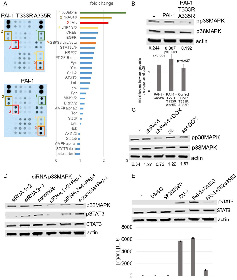
Figure 5. IL-6 and STAT3 activation by PAI-1 in monocytes is p38MAPK-dependent.
A. Phospho-Kinase Array of PB monocytes exposed to rPAI-1 and rPAI-1 T333R A335R (100 nM) for 10 min (left panel). The intensity of the signal was quantified as a fold change in signal intensity in rPAI-1-treated cells over rPAI-1 T333R A335R-treated cells (right panel); B. Top panel: western blot analysis of p38MAPK phosphorylation in lysates of PB monocytes obtained 10 min after exposure to rPAI-1 and rPAI-1 T333R A335R (100 nM). The data are representative of one of 3 independent experiments showing similar results. Lower panel: mean (± 95% CI) fold change in the ratio pp38MAPK:p38MAPK from 3 independent experiments. P values are based on analysis of variance of log10 ratios. C. Western blot analysis of phosphorylation of p38MAPK in lysates of PB monocytes 10 min after exposure to CM from HT1080-Luc-shPAI1 and HT1080-Luc-scPAI1 cultured in the presence or absence of doxycycline. Ratios of pp38MAPK:p38MAPK obtained by scanning densitometry are shown at the bottom of the gel. The data are representative of one of 2 independent experiments showing similar results; D. Western blot analysis of p38MAPK, pSTAT3 and STAT3 in PB monocytes transduced with p38MAPK siRNA and scrambled control siRNA and treated for 7 h with rPAI-1 (100 nM); E. Top panel: western blot analysis of pSTAT3 in PB monocytes exposed for 7 h to 100 nM rPAI-1, DMSO, or SB203580 (20 μM; 1 h preincubation) or their combination. Lower panel: IL-6 protein levels in the medium of PB monocytes under the conditions described in the top panel. The data represent the mean (± SD) of technical duplicates.

