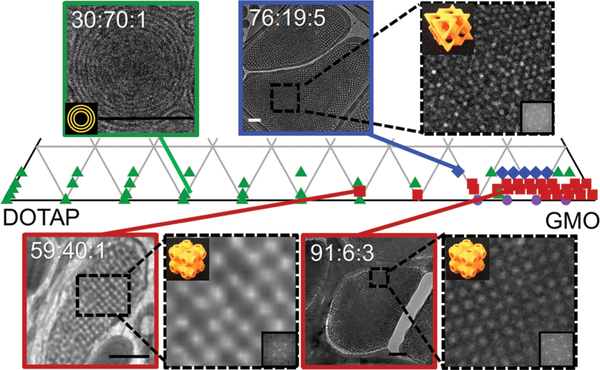
Fig. 4.
Partial phase diagram of GMO/DOTAP/DOPE-PEG in excess water constructed from SAXS data. Multilamellar vesicles (MLV) were observed at low GMO concentration (≤ca. 40 : 60 GMO/DOTAP), and bicontinuous cubic phases were observed at high GMO content (≥ca. 85:15 GMO/DOTAP). Symbols identify structures observed with SAXS: lamellar (green triangle), primitive bicontinuous cubic (red square), diamond bicontinuous cubic (blue diamond), gyroid bicontinuous cubic (purple circle). In some instances, a reentrant lamellar phase is observed at high GMO content. Cryo-EM micrographs are consistent with SAXS data. With 5 mol% DOPE-PEG, diamond bicontinuous cubic phases are observed (e.g., 76 : 19 : 5) and the primitive lattices are preferred for 1 mol% (59 : 40 : 1) and 3 mol% (91 : 6 : 3). Fast Fourier transforms of the enlarged areas (inset) confirm the space group symmetries of the cubic phases (orange 3D models). Scale bars are 200 nm.

