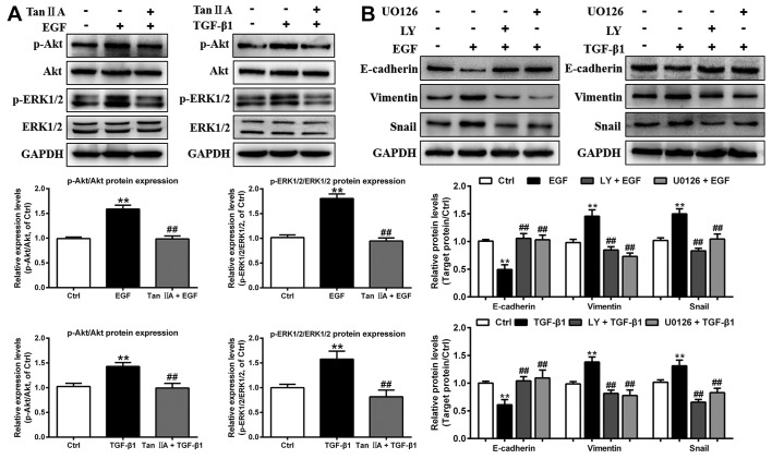
Figure 5.
Tan IIA inhibits EMT by deactivating the PI3K/Akt/ERK signaling pathway in EGF- and TGF-β1-treated HepG2 cells. (A) Phosphorylation and expression of Akt and ERK1/2 in HepG2 cells that were untreated or treated with 20 ng/ml EGF, 10 ng/ml TGF-β1 and 2 µM Tan IIA were analyzed by western blotting. (B) Protein expression levels of E-cadherin, vimentin and Snail in HepG2 cells that were untreated or treated with 20 ng/ml EGF, 10 ng/ml TGF-β1, 20 µM LY, and 20 µM U0126 were analyzed by western blotting. **P<0.01 vs. the control group; ##P<0.01 vs. the EGF or TGF-β1 group. EGF, epidermal growth factor; EMT, epithelial-mesenchymal transition; LY, LY294002; p-, phosphorylated; Tan IIA, tanshinone IIA; TGF, transforming growth factor.

