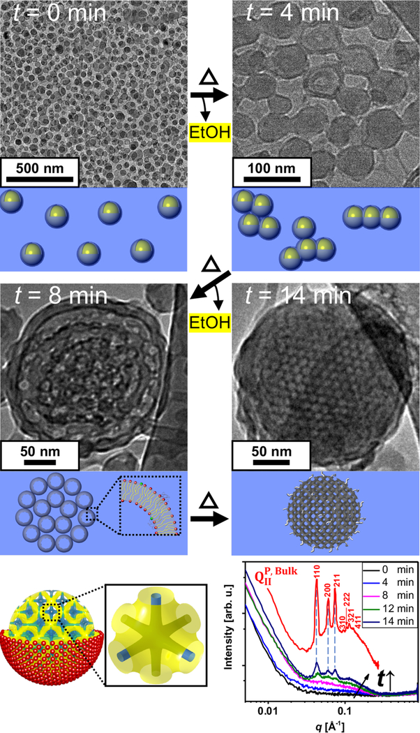
Figure 2.
Mechanism of cubosome formation. Cryo-EM images obtained for microfluidic solutions of ethanol-in-water droplets stabilized by a ternary lipid mixture (GMO/DOTAP/GMOPEG, 97/2/1, mole ratio) at different time points (t) of ethanol evaporation. At t = 0 min monodisperse emulsions (droplet size 50 nm) are observed. At t = 4 min, emulsion droplets begin to fuse into string-like and cluster morphologies. After four more minutes of the treatment (t = 8 min) the membranes in the emulsion are fully fused and starting to display a periodic array. This is the stage where most of nanoparticles show a rim that is consistent with a lipid bilayer structure. At the final stage of the treatment (t = 14 min), lipid membranes now depleted from ethanol rearrange into a periodic and highly ordered primitive bicontinuous cubic structure. Schematics of a cubosome having a primitive bicontinuous cubic structure enclosed by a single lipid leaflet are shown where yellow represents the midplane of the lipid bilayer and blue the water channels. A single unit cell is also represented to the right of the schematic. Synchrotron SAXS scans of the ternary mixture (GMO/DOTAP/GMOPEG, 97/2/1, mole ratio) at different rotary evaporation time points are shown in the bottom right. SAXS scan of a bulk cubic phase with the same lipid composition is included in the plot (red line). The sample treated from 0 and 4 min shows weak scattering due to low lipid concentration. At t = 8 min, a bilayer form factor signal starts to be visible, and at t =12 min three clear Bragg peaks at q = 0.043, 0.061, and 0.075 Å −1 are observed exactly matching the q positions of the bulk phase. The three peaks correspond to the [110], [200], and [211] planes that index to a bicontinuous cubic structure with Im3m space group, QIIP. With two more minutes of treatment (t =14 min), more intense Bragg peaks are observed, indicating that more and highly ordered cubosomes are formed.

