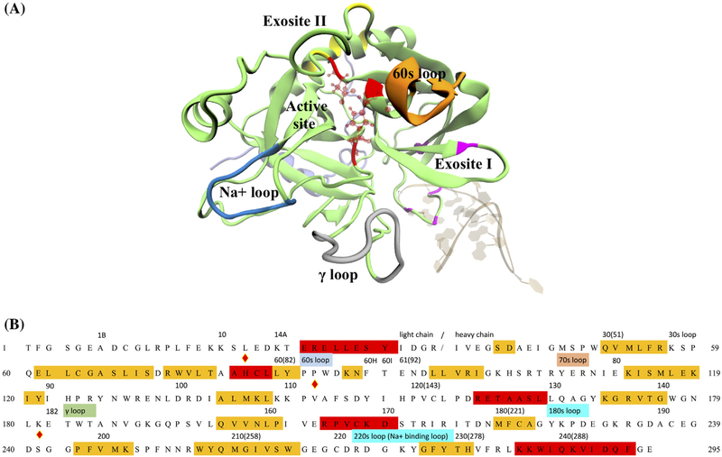
Figure 1.
Human α-thrombin and its functional sites. (A) Tertiary structure of thrombin in PDB 4DII was showed in cartoon representation. The light and heavy chains were respectively colored in light violet and lime. Several known function sites were indicated by the colors and nearby labels. The thrombin-binding aptamer in the same PDB was shown in NewRibbons representation. (B) Sequence of human α-thrombin was listed in one-letter amino acid code. The residue indices in the original PDB file and positions of several function sites were labeled above the sequence. Residues under the red and yellow stand for alpha helix and beta sheet regions. The catalytic triad was marked by the diamond signs above the letter. For the convenience of counting, a new residue number was assigned to each residue as labeled in the beginnings and ends of each row of sequence and the numbers in the parentheses.

