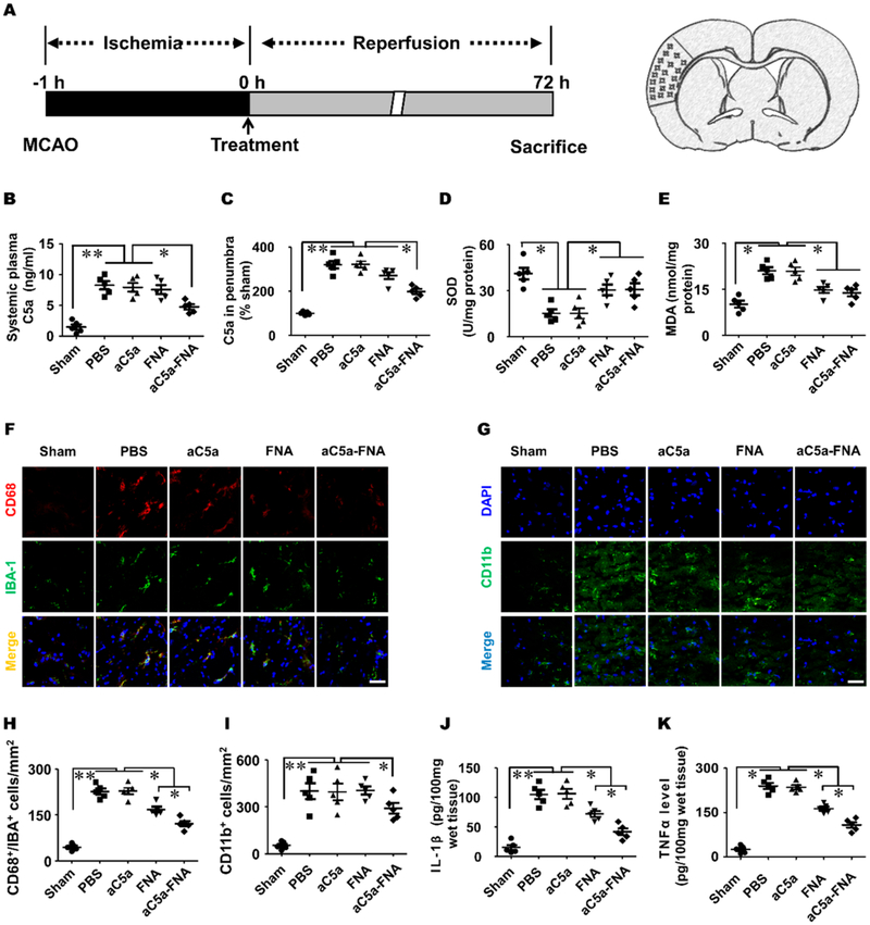
Figure 5.
The aC5a-FNA treatment inhibited oxidative stress and inflammatory in brain penumbra. (A) Time schedule for cerebral IRI establishment and analysis in the ischemic cortex. The change of C5a in (B) the plasma and (C) penumbra. The level of (D) SOD and (E) MDA in the ischemic penumbra (mean ± SD, n = 5, ** P < 0.01). Immunofluorescent staining of (F) CD68+/Iba-1+ and (G) CD11b+ inflammatory cells at 3 days after cerebral IRI. Scale bar: 25 μm. Quantitative analysis of (H) CD68 and Iba-1 double-positive cells and (I) CD11b+ inflammatory cells in the penumbra and (J) IL-1β levels and (K) TNF-α levels in the ischemic penumbra were determined by ELISA assays (mean ± SD, n = 5, ** P < 0.01).

