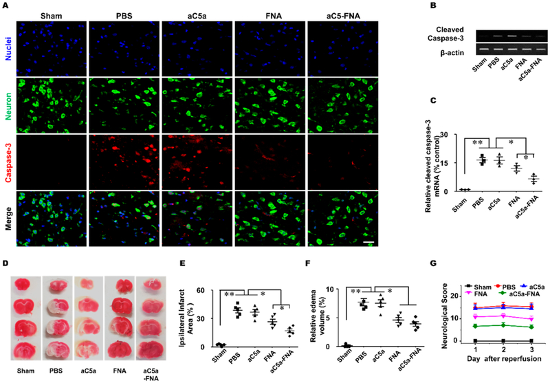
Figure 6.
The aC5a-FNA treatment attenuates brain IRI. (A) Activated caspase-3 (red) and Nissl staining (green) were detected by confocal microscopy in the ipsilateral cortex (scale bar, 25 μm). Activated caspase-3 gene expression in the ischemic penumbra was detected using (B) gel electrophoresis and (C) real-time PCR (mean ± SD, n = 3, ** P < 0.01). (D) Representative TTC staining, quantitative analysis of (E) edema volume, (F) infarct volume, and (G) neurological scores in different groups (n > 3, mean ± SD, *P < 0.05, **P < 0.01).

