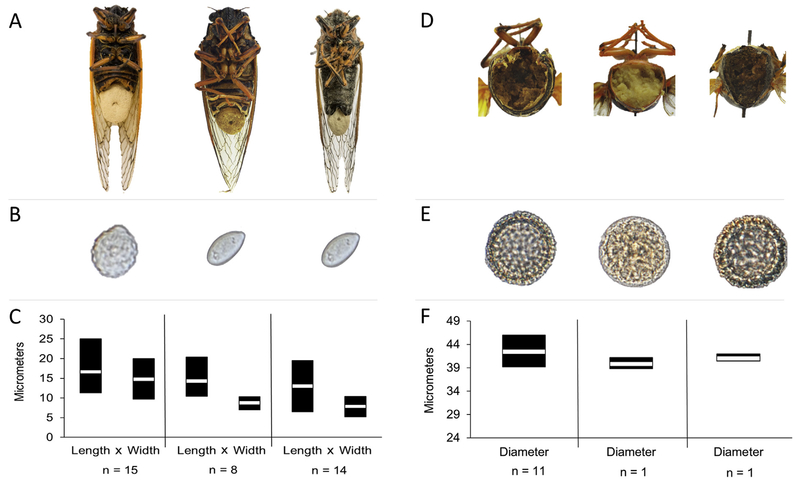
Fig. 1.
Massospora-infected cicadas with associated spore morphology. (A) From left to right: Mas. cicadina-infected periodical cicada (Magicicada septendecim), Mas. levispora-infected Say’s cicada (Okanagana rimosa), and Mas. platypediae infected wing-banger cicada (Platypedia putnami) with a conspicuous conidial “plugs” emerging from the posterior end of the cicada; (B) close-up of conidia for each of three Massospora spp.; (D) posterior cross-section showing internal resting spore infection; and (E) close-up of resting spores for each of three Massospora spp. Specimens in B-F appear in same order as A. Mean (C) conidia and (F) resting spore dimensions for three Massospora species sampled from infected cicadas. Twenty-five conidia or resting spores were measured for each specimen except for Mas. levispora (MI) and Mas. aff. levispora (NM) resting spores, in which 50 spores were measured.

