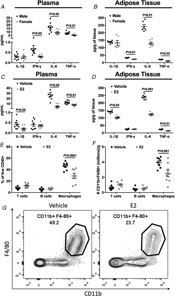
Figure 7. Body weight-matched female mice and oestradiol-treated male mice displayed reduced plasma and adipose tissue inflammation after high fat feeding.
A, plasma IL-1β, IL-6, IFN-γ and TNF-α concentration in male vs. female mice. B, adipose tissue IL-1β, IL-6, IFN-γ and TNF-α concentration in male vs. female mice. C, plasma IL-1β, IL-6, IFN-γ and TNF-α concentration in vehicle vs. oestradiol-treated male mice. D, adipose tissue IL-1β, IL-6, IFN-γ and TNF-α concentration in vehicle vs. oestradiol-treated male mice. E and F, visceral fat immune profile from vehicle vs. oestradiol-treated male mice. G, representative flow cytometry contour plots of adipose tissue macrophages from vehicle vs. oestradiol-treated male mice. Data are means ± SEM.

