
Figure 2.
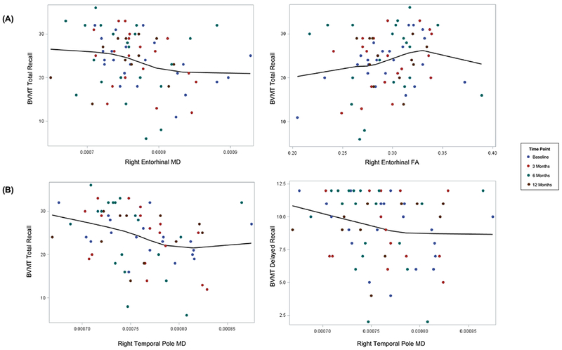
Scatter plots for (A) right entorhinal and (B) right temporal WM and visuospatial memory performance including all time points for each patient up to 12 months post-RT. The trend line overlays the LOESS fit with the smoothing parameter that minimizes the AICC criterion. Significant associations between imaging parameter and memory test were determined based on the beta coefficient (β2) derived from the linear mixed effects model with random intercept and slope:
Raw memory scores are shown. MD is expressed in mm2/s. FA is unitless. Outliers (n=2 and n=4 for A and B, respectively) were removed based on statistically significantly great Mahalanobis distances (P<.001).
(A) Higher right entorhinal MD values were significantly associated with worse BVMT-R Total Recall (β2= −28,385 points/mm2/s, P=.047). Smaller right entorhinal FA values were significantly associated with worse BVMT-R Total Recall (β2= 49.15 points,P=.023). (B) Higher right temporal pole MD values were significantly associated with worse nonverbal memory (BVMT-R Total Recall, β2= −60,800 301 points/mm2/s , P=.003; BVMT-R Delayed Recall, β2= −17,762 301 points/mm2/s, P=.042).
Abbreviations: BVMT-R, Brief Visuospatial Memory Test-Revised; HVLT-R, Hopkins Verbal Learning Test-Revised