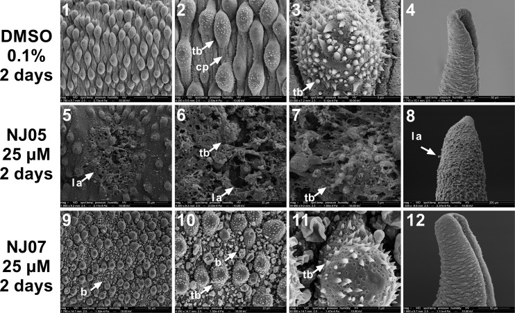
Fig 6. Scanning electron micrographs of dorsal and posterior regions of Schistosoma mansoni adult male worm exposed to NJ05 or NJ07.
Worms were exposed for 2 days to 25 μM NJ05 (panels 5–8), 25 μM NJ07 (panels 9–12) or vehicle (0.1% DMSO) (panels 1–4). Panels 1 to 4: Medial (panels 1–3) and posterior (panel 4) regions of control male worm presented normal tegument (Bar = 50 μm; 20 μm; 5 μm; 50 μm). Note that the size scale bar is shown within the black thin line below each image. Panels 5 to 7: Enlarged view of dorsal region of adult male worm treated with NJ05 showing tegument lesions and loss of tubercles (Bar = 50 μm; 20 μm; 5 μm; 50 μm); panel 8: Posterior region of male adult worm treated with NJ05 showing lesion areas (Bar = 50 μm); panels 9 to 11: Dorsal region of male adult worm treated with NJ07 showed bubbles throughout (Bar = 30 μm; 20 μm; 5 μm; 50 μm); panel 12: Posterior region of male adult worm treated with NJ07 (Bar = 50 μm). cp: ciliated papillae; b: blebs; tb: tubercles; la: lesion area.

