
Fig. 3.
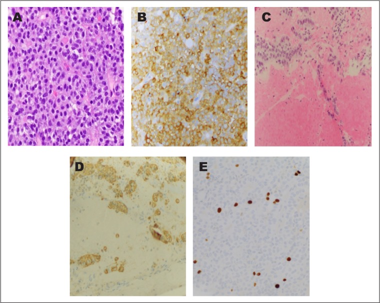
Photomicrographs of the follicle-stimulating hormone-producing adenoma. Monotonous population of tumor cells on hematoxylin and eosin staining (A, ×40) and strong follicle-stimulating hormone immunoreactivity (B, ×20). Pituitary adenoma invading the medial wall of the cavernous sinus (C, ×20) highlighted by an immunostain for chromogranin (D, ×20). A Ki-67 proliferation marker highlights <2% of tumor nuclei (E, ×40).