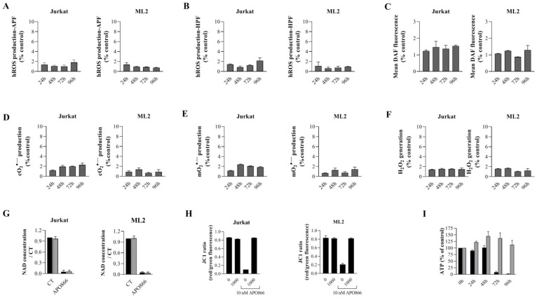
Figure 3. Catalase supplementation abrogates ROS/RNS production, loss of mitochondria membrane potential and ATP, but not NAD depletion in APO866-treated hematological malignant cells.
Time dependent detection of ROS/RNS productions in Jurkat and ML2 cells treated with APO866 (10 nM) in presence of exogenous addition of catalase. Highly reactive ROS (A–B), NO (C), Cytosolic (D), mitochondrial superoxide (E) and hydrogen peroxide (F) were detected as described in Figure 1. Detection of NAD and ATP cell content (G and I), as well as mitochondrial depolarization (H) in APO866-treated Jurkat and ML2 cells in presence (or absence) of catalase (1000 U/ml) were assessed as described in Method section. Total intracellular NAD and ATP contents were measured and normalized to relative protein content and are expressed as a ratio compared to that of untreated Jurkat/ML2 cells.

