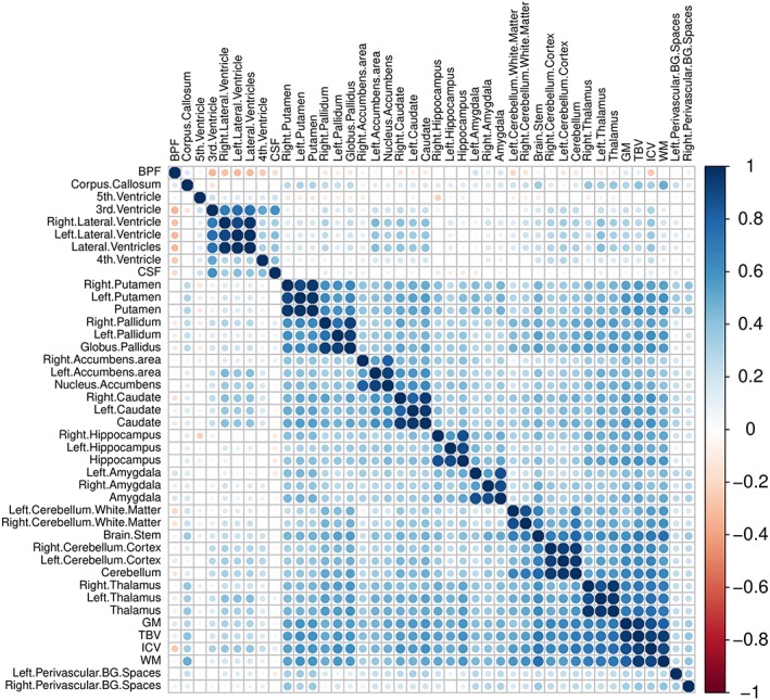
Figure 3.

Correlation heatmap showing pair correlations across brain structures of BREATHE project subsample, with blue color indicating positive correlations and red color indicating negative correlations. 3rd.Ventricle: third ventricle; 4th.Ventricle: fourth ventricle; CSF: cerebrospinal fluid; 5th.Ventricle: septum pellucidum; ICV: total intracranial volume; WM: white matter; TBV: total brain volume; BPF: brain parenchymal fraction; GM: gray matter; Perivascular.BG.Spaces: basal ganglia perivascular spaces. Abbreviations for all brain structures are shown in Table S5
