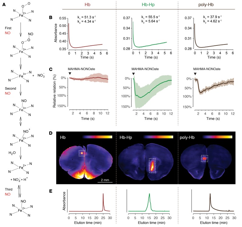
Figure 5. The protective function of Hb-haptoglobin in the brain is determined by its molecular size.
(A) Reaction sequence of oxyHb with NO under conditions of excess NO. In a 3-step reaction, oxyHb can scavenge up to 3 molecules of NO. (B) NO-reaction kinetics of Hb (left), Hb-haptoglobin (middle), or polymerized Hb (right). Shown are absorbance changes at 405 nm after rapid mixing of NO and oxyHb in a stopped-flow spectrophotometer. Estimates of the reaction rates (k1 and k2) were calculated by approximating the data with an equation defined by the sum of 3 exponential functions. (C) Tension traces (mean ± SD) of porcine basilar arteries immersed in buffer containing 10 μM Hb (left; red, n = 16), Hb-haptoglobin (middle; green, n = 16), or polymerized Hb (right, brown, n = 8). A bolus of MAHMA-NONOate was added (arrowhead) to induce NO-mediated dilation. (D) Coronal vibratome sections (2 mm anterior to the bregma) of mouse brains 2 hours after subarachnoid injection of TCO-labeled Hb (left), Hb-haptoglobin (middle) and polymerized Hb (right). The false-colored images represent the signal intensity of the injected compound after postmortem coupling to a fluorophore (tetrazine-5-TAMRA). Intraparenchymal delocalization is only observed in the mouse injected with Hb and not after injection of Hb-haptoglobin and polymerized Hb (images are representative for n = 3 per group). The signal in the area of the puncture channel (dashed boxes) serves as a positive control for the injection and labeling procedure. (E) SEC elution profiles of pure Hb (left; red), Hb-haptoglobin (middle; green), and polymerized Hb (right; brown) measured at 414 nm illustrate the different molecular size of the injected compounds.

