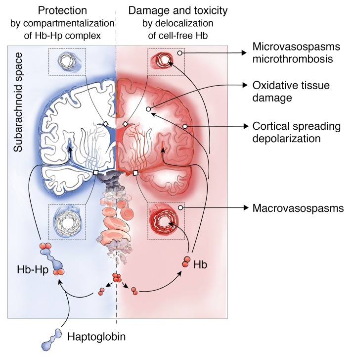
Figure 8. Mechanism of cell-free Hb toxicity and protection by complex formation of Hb with haptoglobin.
Cell-free Hb is released from lysing erythrocytes in the subarachnoid blood clot after aSAH and distributes within the CSF (red). Delocalization of small Hb dimers (32 kDa) into vessel walls of resistance arteries causes macro- and microvascular vasospasms through NO depletion in the vascular wall. Delocalization into the brain parenchyma may cause oxidative damage and alter the interstitial microenvironment. Therapeutic haptoglobin injected into the ventricular system binds Hb in the subarachnoid space and prevents delocalization of the large Hb-haptoglobin complex (>150 kDa), thereby attenuating toxicity (blue).

