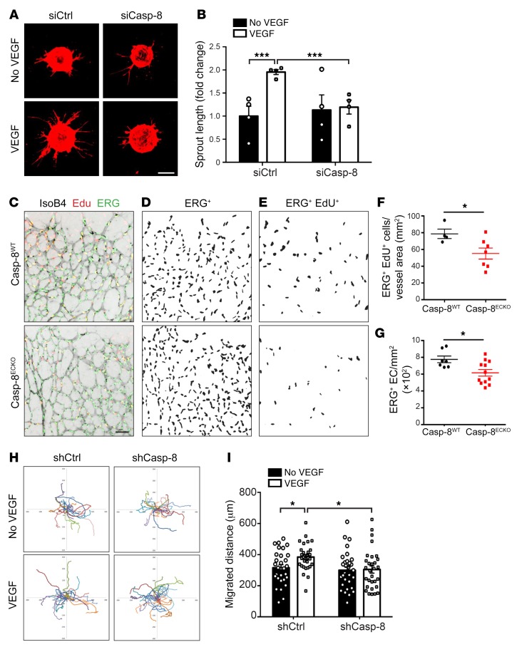
Figure 3. Loss of Casp-8 in ECs impairs sprouting, proliferation, and migration.
(A) Representative images of the bead-sprouting assay using HUVECs transfected with control siRNA (siCtrl) or Casp-8 siRNA (siCasp-8) and treated with VEGF (50 ng/ml) for 24 hours. (B) Quantitative analysis of total sprout length showing that VEGF is not able to induce vessel sprouting in the absence of Casp-8. Approximately 20 beads per condition were quantified. n = 4. (C) Representative images of the retinal vasculature costained with IsoB4, EdU (labels proliferating cells), and ERG (labels EC nuclei) in Casp-8WT and Casp-8ECKO mice. IsoB4 single channel was transformed to gray colors and inverted with ImageJ for better visualization. (D and E) Masks obtained by ImageJ of ERG+ cells (D) and of ERG+ EdU+ proliferating ECs (E). (F and G) Quantification of total number of ECs per retina area (F; n = 7 WT, n = 12 ECKO) and proliferating ECs per vessel area (G; n = 4 WT, n = 7 ECKO), revealing lower absolute EC numbers and fewer proliferating ECs in Casp-8ECKO retinas compared with Casp-8WT littermates. Data from 2 independent litters. (H) Single-cell motility tracks of HUVECs infected with a control (shCtrl) or Casp-8 shRNA lentivirus (shCasp-8) and treated with VEGF (50 ng/ml) for 12 hours. Migration origin of each cell was overlaid at the zero-crossing point. (I) Quantification of the total migration distance of HUVECs in H showing that VEGF-induced migration was impaired in Casp-8KD ECs. At least 30 cells per condition were quantified. n = 3. For B, F, G, and I, data are shown as mean ± SEM. *P < 0.05; ***P < 0.001, 2-way ANOVA with Bonferroni’s multiple comparisons test (B and I); *P < 0.05, 2-tailed unpaired Student’s t test (F and G). Scale bars: 200 μm (A); 50 μm (C).

