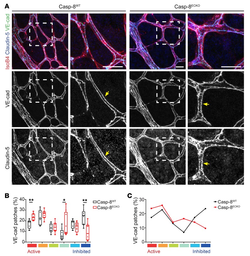
Figure 4. Casp-8 is necessary for maintaining EC junction stability at the retina plexus in vivo.
(A) Representative images of Casp-8WT and Casp-8ECKO retinas stained with IsoB4, VE-cadherin, and claudin-5 (insets of left panels are shown on the adjacent right panels), showing that junctions are more serrated and discontinuous at the retina plexus in Casp-8ECKO compared with Casp-8WT mice (yellow arrows). Images of VE-cadherin and Claudin-5 single channels were transformed to gray colors with ImageJ for better visualization. (B) Quantification of the percentage of VE-cadherin patches showing a significant increase in the number of highly active VE-cadherin patches and a lower number of highly inhibited patches in Casp-8ECKO compared with Casp-8WT mice. Each box shows the median percentage of patches of that type (line) and upper and lower quartiles (box). The whiskers extend to the most extreme data within 1.5 times the interquartile range of the box. *P < 0.05; **P < 0.01, Dirichlet regression model with 2-tailed Mann-Whitney U test for each state. n = 9 WT, n = 9 ECKO. (C) Average of the differential distribution of the percentage of VE-cadherin patches in Casp-8WT and Casp-8ECKO retinas. Scale bars: 20 μm (A).

