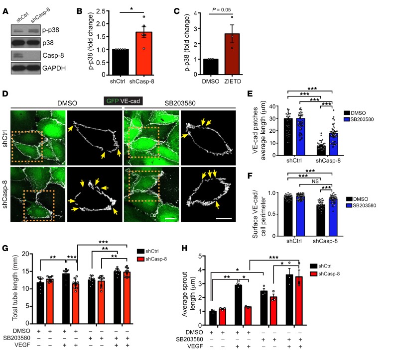
Figure 6. Loss of Casp-8 results in basal activation of p38 MAPK, VE-cadherin instability, and defects in angiogenesis in vitro.
(A)Western blots showing increased p-p38 under basal conditions in Casp-8KD (shCasp-8) ECs compared with control. (B) Quantification of p-p38 as in A. n = 5. (C) Quantification of p-p38 of HUVECs treated with ZIETD (10 μM, 16 hours) under basal conditions, showing that blocking Casp-8 activity also induces increased basal p-p38. n = 3. (D) Images of VE-cadherin staining in shCtrl and shCasp-8–infected HUVECs treated with or without p38 inhibitor (SB203580, 1 μM, 16 hours). Yellow arrows point to empty VE-cadherin spots. (E) Quantification of VE-cadherin average patch length from cells as in H. (F) Quantification of the total amount of VE-cadherin per cell perimeter reveals that inhibition of p38 (SB203580) in Casp-8KD ECs restores VE-cadherin to control levels. At least 15 cells per condition were quantified. n = 3. (G) Quantification of total tube length of HUVECs treated as in E showing that blocking p38 in Casp-8KD ECs restores VEGF-induced tube formation. 3 fields per condition were quantified. n = 4. (H) Quantitative analysis of total sprout length showing that inhibition of p38 rescues VEGF-induced EC sprouting in Casp-8KD ECs. Approximately 20 beads per condition were quantified. n = 3. For B, C, and E–H, data represent mean ± SEM. *P < 0.05, 1-sample t test (B and C); *P < 0.05; **P < 0.01; ***P < 0.001, 2-way ANOVA with Bonferroni’s multiple comparisons test (E–H). Scale bars: 20 μm (D).

