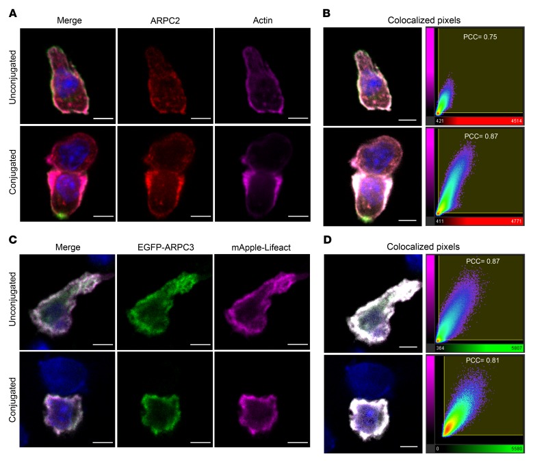
Figure 1. Arp2/3 colocalizes with actin in migrating and synapse-forming CTLs.
(A) Single confocal slices of untransfected OT-I CTLs stained with antibodies against CD8 (green), ARPC2 (red), and actin (magenta). (B) Left panels: merged images from A illustrating the colocalized regions in white saturation after analysis. Right panels: colocalization graphs of ARPC2 voxels (red x axes) plotted against actin voxels (magenta y axes). (C) Single confocal slice of OT-I CTLs transfected with EGFP-ARPC3 (green) and mApple-Lifeact (magenta) constructs. (D) Left panels: merged images from C showing the colocalized regions in white saturation after analysis. Right panels: colocalization graphs of EGFP-ARPC3 voxels (green x axes) plotted against mApple-Lifeact voxels (magenta y axes). Conjugated cells were fixed 25 minutes after mixing with OVA-loaded EL4 target cells. Numbers on the graphs indicate the degree of colocalization expressed as a PCC. Nuclei stained with Hoechst (blue). Scale bars: 4 μm. Data representative of 2 independent experiments. (A) CTLs = 75; conjugates = 48. (C) CTLs = 66; conjugates = 44).

