
Fig. 2 a.
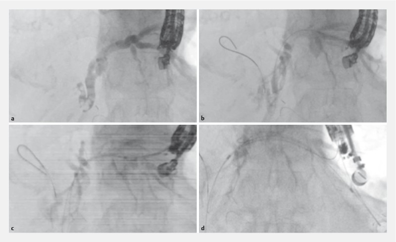
The contrast medium is injected after the intrahepatic bile duct is punctured using a 19G needle. b The 0.025-inch guidewire is inserted into the biliary tract. c The bile duct wall is dilated using the novel electrocautery dilator. d Covered self-expandable metal stent deployment is performed from the intrahepatic bile duct to the stomach.